
What is an angiography?
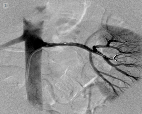
An angiography is a type of X-ray which examines the blood vessels.

What happens during an angiography?
First, a catheter is inserted into one of your arteries, depending on the area which needs to be examined. Through this catheter, a dye is administered to contrast the area that needs to be visualised. The catheter is then guided through the arterial system to the area which needs to be examined. X-rays are then taken as the contrast dye flows through the blood vessels.
Why is an angiography performed?
An angiography is done to check how the blood flows through the vessels, and the general health of the blood vessels too. An angiography is useful in investigating and diagnosing problems affecting the blood vessels, such as narrowing of the arteries, blood clots, or embolisms.
Preparing for angiography
Before the test, the patient is examined, including a blood test. The clinic or doctor will ask you about your medical history and if you are taking any medications. The doctor will also ask if you would prefer to be sedated during the procedure.
What does the exam feel like?
The X-ray table can be somewhat uncomfortable, and the procedure generally lasts between 30 minutes and two hours. You may also feel a pinch when local anaesthesia is administered, but the area is generally numbed so it shouldn’t hurt. When the catheter is inserted and moved around the body, you may feel a pushing or pulling sensation, but it should not be painful.
What happens after the test?
After the angiography, you will be moved to an area where you can rest, and you have to lie still for a few hours to prevent bleeding. Generally angiographies are performed as a day case procedure, although in some cases you may be kept in overnight for monitoring. Generally results of the test are available after a few weeks. You may feel some discomfort, such as soreness at the entry site for a few days after the procedure. Some bruising may also occur.
